
Protocol for dental diafanization
Article Sidebar
Main Article Content
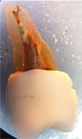
Background: Dental diaphonization is a technique that allows the teeth to be made transparent, making the internal anatomy of the root canals visible, offering an economical and reliable pedagogical tool; however, the literature does not report standardized protocols to obtain a predictable dental clearance.
Aim: obtain a standardized protocol for dental diaphonization as an educative model from the review of the literature and the realization of a pilot study.
Materials and methods: A systematic search was made on databases Scopus and Medline, with the Mesh terms "root canal", "diaphonization", "clearing", "morphology" and, “anatomy", and an extraction table was structured with the most representative variables to establish the three diaphanization phases, a. Decalcification, 5% Nitric Acid, 10% Formic Acid (TBD-2) and 10% EDTA were evaluated, b. Dehydration, ascending Ethyl Alcohols were used, c. Clarification, Methyl Salicylate, and Immersion synthetic oil were evaluated. 54 teeth were selected, 36 without root canal treatment and 18 with root canal treatment, then they were distributed into two groups: Group A, Teeth without root canal treatment, and Group B, Teeth with root canal treatment. Each group was constituted of 18 subgroups defined in order of the decalcification agent type, moment of the contrast medium application, and clarification agent type.
Results: Even though 5% Nitric Acid was the most corrosive agent, it allowed a better flow and accessibility for the contrast medium (Chinese ink) in teeth without root canal treatment. Likewise, 10% Formic Acid preserved the structure of the endodontic tooth. As a clarification agent, the Methyl Salicylate showed better visual results, achieving greater transparency.
Conclusion: The development of a pilot study aimed to standardize diaphonization techniques in dentistry allows the structuring of educative protocols that permit knowing the great tooth anatomic variability and the comprehension as well as the analysis of the root canal treated teeth, contributing to a pedagogic tool for the root anatomy awareness. Using 10% Formic Acid on root canal-treated teeth and 5% Nitric Acid on root canal not treated teeth, with transparency achieved by using Methyl Salicylate, showed better visual results on anatomy and endodontic filling.
Versiani MA, Basrani B, Sousa-Neto MD. The root canal anatomy in permanent dentition. The Root Canal Anatomy in Permanent Dentition. 2018. 1–425 p.
Neelakantan P, Subbarao C, Subbarao C V. Comparative Evaluation of Modified Canal Staining and Clearing Technique, Cone-Beam Computed Tomography, Peripheral Quantitative Computed Tomography, Spiral Computed Tomography, and Plain and Contrast Medium–enhanced Digital Radiography in Studying. J Endod [Internet]. 2010 Sep 1;36(9):1547–51. Available from: https://doi.org/10.1016/j.joen.2010.05.008
Parekh V, Shah N, Joshi H. Root canal morphology and variations of mandibular premolars by clearing technique: An in vitro study. J Contemp Dent Pract. 2011;12(4):318–21.
Robertson DC, Leeb J. The evaluation of a transparent tooth model system for the evaluation of endodontically filled teeth. J Endod. 1982;8(7):317–21.
Venturi M, Prati C, Capelli G, Falconi M, Breschi L. A preliminary analysis of the morphology of lateral canals after root canal filling using a tooth-clearing technique. Int Endod J. 2003;36(1):54–63.
Black G V. Descriptive anatomy of the human teeth [Internet]. 1902. Available from: https://www.worldcat.org/title/descriptive-anatomy-of-the-human-teeth/oclc/605487829
von Lunkaszprie GC. Anatomie des Mundes [Internet]. Braunmüller und Seidel; 1842. (Systematisches Handbuch der Zahnheilkunde). Available from: https://books.google.com.co/books?id=SFgPzQEACAAJ
Paul de Terra. Vergleichende anatomie des menschlichen gebisses und der zähne der vertebraten, [Internet]. 1911. Available from: https://www.worldcat.org/title/vergleichende-anatomie-des-menschlichen-gebisses-und-der-zahne-der-vertebraten/oclc/9704460
Goldman M, Pearson AH, Darzenta N. Endodontic success—Who’s reading the radiograph? Oral Surgery, Oral Med Oral Pathol [Internet]. 1972 Mar 1 [cited 2020 Mar 27];33(3):432–7. Available from: https://www.sciencedirect.com/science/article/abs/pii/0030422072904732?via%3Dihub
Omer OE, Shalabi RM Al, Jennings M, Glennon J, Claffey NM. A comparison between clearing and radiographic techniques in the study of the root‐canal anatomy of maxillary first and second molars. Int Endod J [Internet]. 2004; Available from: https://onlinelibrary.wiley.com/doi/full/10.1111/j.0143-2885.2004.00731.x
Baumann MA, Doll GM. Spatial reproduction of the root canal system by magnetic resonance microscopy. J Endod [Internet]. 1997 Jan 1;23(1):49–51. Available from: https://doi.org/10.1016/S0099-2399(97)80207-5
Green EN. Microscopic investigation of root canal diameters. J Am Dent Assoc [Internet]. 1958 Nov 1;57(5):636–44. Available from: https://doi.org/10.14219/jada.archive.1958.0254
Skidmore AE, Bjorndal AM. Root canal morphology of the human mandibular first molar. Oral Surgery, Oral Med Oral Pathol [Internet]. 1971 Nov 1 [cited 2020 Mar 27];32(5):778–84. Available from: https://www.sciencedirect.com/science/article/abs/pii/0030422071903045?via%3Dihub
Kartal N, Özçelik B, Cimilli H. Root canal morphology of maxillary premolars. J Endod [Internet]. 1998 Jun 1 [cited 2020 Mar 27];24(6):417–9. Available from: https://www.sciencedirect.com/science/article/pii/S0099239998800241
Durack C, Patel S. Cone beam computed tomography in endodontics. Braz Dent J [Internet]. 2012 [cited 2020 Mar 2];23(3):179–91. Available from: http://www.scielo.br/scielo.php?script=sci_arttext&pid=S0103-64402012000300001&lng=en&tlng=en
B. C. W. Barker B. C. Lockett K. C. Parsons. The demonstration of root canal anatomy. Aust Dent J. 1969;
O’Neill KJ, Pitts DL, Harrington GW. Evaluation of the apical seal produced by the McSpadden Compactor and by lateral condensation with a chloroform-softened primary cone. J Endod [Internet]. 1983 May 1;9(5):190–7.
Kasahara E, Yasuda E, Yamamoto A, Anzai M. Root canal system of the maxillary central incisor. J Endod [Internet]. 1990 Apr 1;16(4):158–61.
Saunders WP, Saunders EM. Effect of noncutting tipped instruments on the quality of root canal preparation using a modified double-flared technique. J Endod [Internet]. 1992 Jan 1;18(1):32–6.
Tagger M, Katz A, Tamse A. Apical seal using the GPII method in straight canals compared with lateral condensation, with or without sealer. Oral Surgery, Oral Med Oral Pathol [Internet]. 1994 Aug 1 [cited 2020 Mar 27];78(2):225–31.
Felton DA, Webb EL, Kanoy BE, Dugoni J. Threaded endodontic dowels: Effect of post design on incidence of root fracture. J Prosthet Dent [Internet]. 1991 Feb 1;65(2):179–87. Available from: https://doi.org/10.1016/0022-3913(91)90159-T
Vertucci FJ. Root canal morphology of mandibular premolars. J Am Dent Assoc [Internet]. 1978 Jul 1;97(1):47–50. Available from: https://doi.org/10.14219/jada.archive.1978.0443
Okumura T. Anatomy of the Root Canals. J Am Dent Assoc [Internet]. 1927 Apr 1 [cited 2020 Mar 27];14(4):632–6. Available from: https://www.sciencedirect.com/science/article/abs/pii/S1048636427440093?via%3Dihub
Greco Machado, Y., García Molina, J. A., Bueno Martínez, R., Manzanares Céspedes, M. C., & Lozano de Luaces, V. (2008). Técnicas de diafanización: estudio comparativo. Endodoncia, 2008, vol. 26, num. 2, p. 85-92.
Pediatrica E, Al R, Julian E, Preliasco M. Validación de modelos de simulación para práctica pre clínica en Endodoncia pediatrica. Resistencia al degaste. 2018;
Andrés MGS. “Estudio in-vitro de la anatomía interna de conductos radiculares del primer premolar superior, estudio mediante la técnica de diafanización dental.” 2018.
Rehman K, Khan FR, Habib S. Diaphonization: a recipe to study teeth. J Contemp Dent Pract. 2015 Mar 1;16(3):248-51. doi: 10.5005/jp-journals-10024-1670. PMID: 26057927.
Chin, R. L., Olson, K. R., & Dempsey, D. (2007). Salicylate toxicity from ingestion and continued dermal absorption. The California journal of emergency medicine, 8(1), 23–25.
Beatriz Labarta, A., Cuadros, M., Gualtieri, A., & Sierra, L. (2016). Evaluación de la morfología radicular interna de premolares inferiores mediante la técnica de diafanización, obtenidos de una población argentina. Revista Científica Odontológica, 12.
Vertucci FJ. Root canal anatomy of the human permanent teeth. Oral Surg Oral Med Oral Pathol. 1984 Nov;58(5):589-99. doi: 10.1016/0030-4220(84)90085-9. PMID: 6595621.
Pécora JD, Woelfel JB, Sousa Neto MD, Issa EP. Morphologic study of the maxillary molars. Part II: Internal anatomy. Braz Dent J. 1992;3(1):53-7. PMID: 1303118.
Castelucci A. Endodontics. Edizioni Odontoiatriche Il Tridente: Florence; 2005
Castania VA, Silveira JW, Issy AC, Pitol DL, Castania ML, Neto AD, Bel EA, Defino HL. Advantages of a combined method of decalcification compared to EDTA. Microsc Res Tech. 2015 Feb;78(2):111-8. doi: 10.1002/jemt.22451. Epub 2014 Nov 28. PMID: 25452153.
Choube A, Astekar M, Choube A, Sapra G, Agarwal A, Rana A. Comparison of decalcifying agents and techniques for human dental tissues. Biotech Histochem. 2018;93(2):99-108. doi: 10.1080/10520295.2017.1396095. Epub 2018 Jan 9. PMID: 29313383.
Sanjai K, Kumarswamy J, Patil A, Papaiah L, Jayaram S, Krishnan L. Evaluation and comparison of decalcification agents on the human teeth. J Oral Maxillofac Pathol. 2012 May;16(2):222-7. doi: 10.4103/0973-029X.99070. PMID: 22923894; PMCID: PMC3424938.
Gupta, S., Jawanda, M. K., Sm, M., Bharti, A. (2014). Qualitative histological evaluation of hard and soft tissue components of human permanent teeth using various decalcifying agents - a comparative study. Journal of clinical and diagnostic research: JCDR, 8(9), ZC69–ZC72. https://doi.org/10.7860/JCDR/2014/10195.4874
Robertson D, Leeb IJ, McKee M, Brewer E. A clearing technique for the study of root canal systems. J Endod. 1980 Jan;6(1):421-4. doi: 10.1016/S0099-2399(80)80218-4. PMID: 7005366.
Armarego WLF. Purification of laboratory chemicals. Purif Lab Chem. 2017;1–1176.
- Ivan Felipe Restrepo Salas, Gilbert Alfonso Morales , Ingrid Ximena Zamora , Carlos Humberto Martínez , Anatomy of the pulp chamber and the root canal system: Pedagogical strategies A literature review , Revista Estomatología: Vol. 31 No. 2 (2023)


Accepted 2023-03-06
Published 2023-10-06

This work is licensed under a Creative Commons Attribution-NonCommercial-NoDerivatives 4.0 International License.
Los autores/as conservan los derechos de autor y ceden a la revista el derecho de la primera publicación, con el trabajo registrado con la licencia de atribución de Creative Commons, que permite a terceros utilizar lo publicado siempre que mencionen la autoría del trabajo y a la primera publicación en esta revista.